Researchers from Sungkyunkwan University (SKKU) in South Korea, in collaboration with colleagues from the United States, have introduced a technology that could mark a turning point in orthopedic surgery: a modified glue gun that prints biodegradable bone grafts directly onto fractures and defects during surgery. The advancement, published in Cell Press’s Device journal, combines low-temperature 3D printing, next-generation biomaterials, and a portable design tailored for the operating room.
From Household Tool to Medical Device
The team transformed a common glue gun into a real-time in situ printing system capable of extruding biomaterials at temperatures safe for human tissue (around 60°C). The result is a compact, hand-held, easy-to-handle device that allows the surgeon to control the direction, angle, and depth of printing in real time.
This represents a shift from current practice, which requires manufacturing custom implants before surgery through medical imaging, 3D modeling, and laboratory processes. With this technique, the graft is created directly on the damaged bone within minutes.

A Material Designed to Heal and Disappear
The “sticks” in the gun do not contain glue but a blend of polycaprolactone (PCL) and hydroxyapatite (HA):
- PCL is a biodegradable polymer that offers mechanical strength and elasticity.
- HA is a natural mineral found in bone that enhances cell adhesion and stimulates new bone tissue growth.
The resulting scaffold performs three key functions:
- Biologically integrates with the surrounding healthy bone.
- Serves as a temporary support while new tissue grows.
- Degrades in a controlled manner, being replaced by regenerated bone.
Furthermore, researchers demonstrated that adding antibiotics like vancomycin or gentamicin into the mix allows the graft to release drugs locally over weeks, reducing the risk of postoperative infections and the reliance on oral treatments.
Laboratory and Animal Model Results
The team validated the material through several phases:
- Cell culture: confirming the compound is non-toxic and promotes adhesion and proliferation of bone cells.
- Mechanical testing: measuring resistance to compression, bending, bonding to bone, and degradation rate.
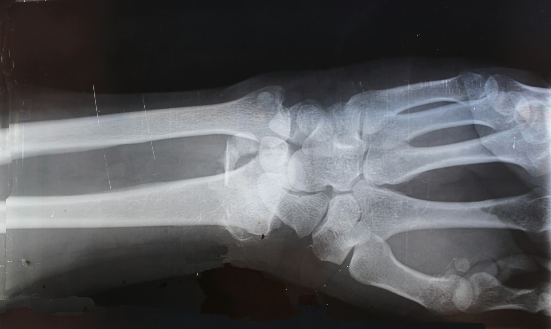
- Animal model: in rabbits with femoral defects too large to heal on their own, comparing the printed bone grafts to commercial bone cement.
Over a 12-week period, the printed grafts showed:
- Better bone regeneration with a more natural structure.
- Higher bone surface area and cortical thickness (indicators of strength).
- No signs of inflammation or necrosis in surrounding tissues.
- Significant antibacterial effect when antibiotics were added.
Advantages Over Traditional Methods
Currently, bone defects are treated with:
- Commercial bone cements, which seal but do not promote regeneration well.
- Bone grafts, requiring tissue extraction from another site or donors.
- Metal implants, effective but costly and less adaptable to irregular defects.
The bone gun offers an alternative that is:
- More personalized: directly prints onto irregular fractures.
- Faster: the process completes in minutes, reducing surgical time.
- More cost-effective: avoids external manufacture of implants.
- Safer: reduces infections by locally releasing antibiotics.
Challenges Toward Clinical Implementation
Though promising, the technology is still far from application in humans. The researchers acknowledge several remaining steps:
- Sterilization protocols for the device and the material.
- Trials in larger animals to demonstrate efficacy and safety.
- Standardization of production of the biomaterial sticks.
- Regulatory approvals needed for large-scale medical use.
Future Implications
If these stages are successfully completed, the bone printing gun could revolutionize orthopedics and traumatology, with applications beyond fractures:
- Reconstruction after bone cancer.
- Correction of congenital defects.
- Personalized regenerative medicine, incorporating various drugs or materials tailored to each patient.
Additionally, the concept of in situ biomedical printing opens doors to other uses: printing cartilage, skin, or even soft tissue during reconstructive surgeries.
Conclusion
The bone gun developed in South Korea represents a disruptive potential advancement: a portable device that turns 3D printing into a direct surgical tool, efficient and adaptable. Although it still needs to overcome regulatory testing and human trials, laboratory and animal model results suggest that it could become the future standard for rapid, personalized bone repair.
Frequently Asked Questions
What materials does the bone printing gun use?
A composite of polycaprolactone (PCL), a biodegradable polymer, and hydroxyapatite (HA), a natural mineral found in bone, with the option to add antibiotics.
Why is it better than bone cement?
Because it not only seals but also stimulates natural bone regeneration, integrates with surrounding tissue, and gradually disappears as new bone forms.
What clinical applications does it have?
Complex fractures, post-tumor resection reconstructions, congenital defects, or orthopedic surgeries requiring custom implants.
When might it be available for human patients?
Animal trials in larger models, sterilization protocols, and regulatory approvals are still needed. If these are successful, it could reach operating rooms in a few years.
via: mentes curiosas, scimex and Cell